Masking
For each image slice in the Visible Human Project, a “mask slice” was created. Mask slices are black-and-white (0-1) binary images, where the white pixels represent data to be processed and the black pixels represent data to be filtered out, i.e., irrelevant features in the images such as the blue background from the dyed gelatin solution fluid [1]. For example, here is the mask slice 1108 that corresponds to the image slice 1108. The mask slices were first created by using a thresholding algorithm on the color image slices (in the L*a*b* color space [5]) to separate the blue background from the body [6]. This automated approach resulted in inaccurate masks because of several reasons related to the nature of the images and the method used to obtain them in the Visible Human Project:
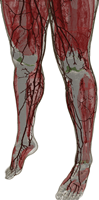
- Two Tygon tubes (3-mm outer diameter) filled with contrast agents for the CT and MR were attached to the body to serve as fiduciary markers between the different data sets [1]. The tubes, which run along the skin surface of the anterior aspect from the head down to each foot, are a different color than the blue background as highlighted here for the image slice 1108.
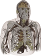
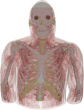
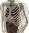
- In some image slices, the body parts underneath the slice are partially visible through the dyed gelatin solution. This causes the gelatin solution’s color to be similar to skin color in places where the body expands in the slices underneath current slice (for example, the shoulders are broader than the neck and are visible in the slices above them as shown here for the image slice 1242).
- Some of the body cavities were filled with the dyed gelatin solution (such as the ear canal) while other cavities were filled with latex to prevent debris from collecting in the cavities during the preparation of the body. Both the dyed gelatin solution and the latex are blue in the images slices [1]. This requires a careful identification of the boundaries of these cavities (see the segmentation step) to avoid filtering out important body features. This identification step can be delayed by filling in the cavities in the mask slices, e.g., see the ear canals in the image slice 1135 and its mask slice 1135.
- Other odd protrusions from the skin also appeared in some of the pictures where the background was lighter in color due to the intentionally conservative choice of the thresholds. If the odd protrusions were not fixed in the masks, then they would appear in the homogeneous model that is derived from the masks, even though the odd protrusions could later be labeled as air in the heterogeneous model. These protrusions can be seen in the uncorrected mask slice 1009 and are removed (using 1. below) in the final mask slice 1009, where both of these examples were zoomed in on to show the detail.
- Some of the image slices are severely degraded or missing because of the saw kerf that occurred during the preparation of the body for the cross-sectional slices [1]. The body was cut into 4 segments using a high-tension backsaw so that each segment would fit inside they cryomacrotome. These cuts affect slices 1505 - 1507, 2016 - 2022, and 2424 - 2435.
These complications were addressed by editing the mask slices as follows:
- The bumps in the images from the fiduciary markers and the odd protrusions from the skin were removed by dilating and then eroding (using a disk-shaped structuring element with a radius of 5 pixels) the white regions [7].
- The resulting black-and-white masks were individually reviewed for any discrepancies. These discrepancies were corrected by displaying the outline of the initial mask on top of the original image in the background, manually selecting problem pixels, and assigning them to either the foreground or background.
- The degraded or missing image slices were replaced with the closest valid slice. In effect, the closest valid slices were extruded further and the z-resolution of the model decreased at slices 1505 - 1507, 2016 - 2022, and 2424 - 2435.
The mask slices are combined with the image slices (using the logical “and” operation at each pixel) to prepare them for the next step. This operation simplifies and speeds up the segmentation step because it reduces the image/data size and filters out irrelevant features. Moreover, the mask slices can be used to create homogeneous voxel models; however, because of the above listed complications, the white regions in most of the mask slices do not contain any cavities and are by design (slightly) larger than the regions of interest, i.e., the resulting homogenous voxel model is larger than the actual body.
| Previous: Cropping | Up: Methodology | Next: Segmentation and Material Identification |
