Image Shifts
The cross-sectional image slices in the Visible Human Dataset do not align perfectly as observed in [12]. At some of the image slices, the picture is significantly shifted compared to the one above it and thus all the slices below that are misaligned by that shift (Fig. 4). To counter the misalignment, the slices where these shifts occur are identified, the amount of the shift is determined, and the slices are shifted back. The shifts were quantized in terms of pixels and their values were determined by maximizing the overlap between the mask of the slices under operation and the slices above them (e.g., mask of slice 1231 was compared to that of slice 1230). The shifts were then confirmed by visual inspection. Table 1 shows the pixel shifts made to the slices while developing the AustinMan model.
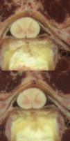
Figure 4: A cropped portion (at the same position of each slice) of the
spinal cord from slices a_vm1230.png (top) and a_vm1231.png (bottom)
indicating the relative shift of 9 pixels to the right.
Table 1: Pixel Shifts Used to Align the Dataset
| Relative Pixel Shift to the Right (in the x-direction) Compared to Previous Image | Cumulative Pixel Shifts to the Right (in the x-direction) |
| Slice 1231: 9 | Slices 1231-1252: 9 |
| Slice 1253: -11 | Slices 1253-1360:-2 |
| Slice 1361: 13 | Slices 1361-1390: 11 |
| Slice 1391: -13 | Slices 1391-1416: -2 |
| Slice 1417: 13 | Slices 1417-1729: 11 |
| Slice 1730: 6 | Slices 1730-1790: 17 |
| Slice 1791: 1 | Slices 1791-1880: 18 |
| Slice 1881: -2 | Slices 1881-2546: 16 |
| Slice 2547: 2 | Slices 2547-2702: 18 |
| Slice 2703: 2 | Slices 2703-2878: 20 |
| Previous: Assumptions | Up: Assumptions | Next: Tissue Material Assumptions |
